Use code RJTJFG for 20% off at checkout. Until 5/31/20

Boundary: Bleed area may not be visible.

The watermark at the lower right corner of the image will not appear on the final product.

by Living Art Enterprises and Photo Researchers
$54.00
This product is currently out of stock.
Size
Orientation
Image Size
Product Details
You'll never run out of power again! If the battery on your smartphone or tablet is running low... no problem. Just plug your device into the USB port on the top of this portable battery charger, and then continue to use your device while it gets recharged.
With a recharge capacity of 5200 mAh, this charger will give you 1.5 full recharges of your smartphone or recharge your tablet to 50% capacity.
When the battery charger runs out of power, just plug it into the wall using the supplied cable (included), and it will recharge itself for your next use.
Design Details
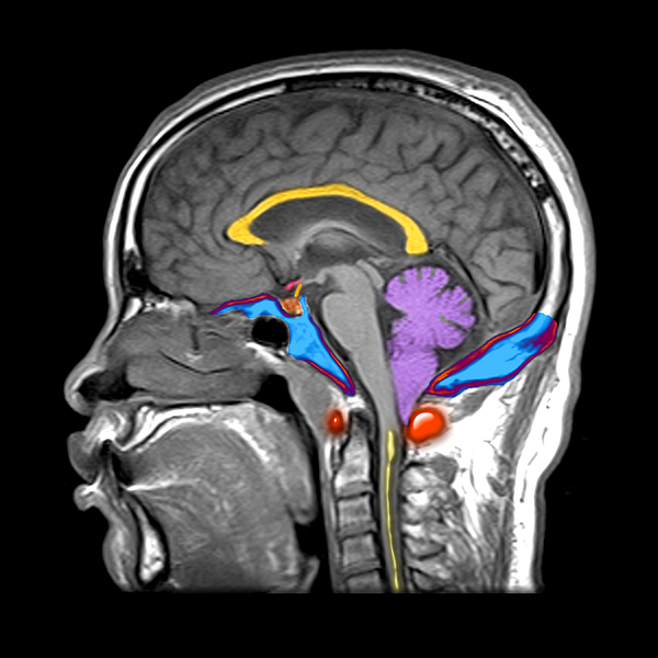
Color enhanced sagital MRI of the brain of a person with a Chiari I malformation. This is a congenital anomaly in the development of the hindbrain... more
Dimensions
1.80" W x 3.875" H x 0.90" D
Ships Within
1 - 2 business days

Photograph

Canvas Print

Framed Print

Art Print

Poster

Metal Print

Acrylic Print

Wood Print

Greeting Card

iPhone Case

Throw Pillow

Duvet Cover

Shower Curtain

Tote Bag

Round Beach Towel

Zip Pouch

Beach Towel

Weekender Tote Bag

Portable Battery Charger

Bath Towel

Apparel

Coffee Mug

Yoga Mat

Spiral Notebook

Fleece Blanket

Tapestry

Jigsaw Puzzle

Sticker
Color enhanced sagital MRI of the brain of a person with a Chiari I malformation. This is a congenital anomaly in the development of the hindbrain (posterior fossa). The corpus callosum (yellow), the pituitary gland (small rounded brown structure with a stem), the cerebellum (purple), and the cerebellar tonsil (purple peg like structure projecting into the upper cervical spinal canal) are visible. The blue structures with red rim are the bones of the skull base and the bright red structure is the first cervical vertebrae (the atlas). The vertical yellow stripe represents a syrinx (cavity) in the cervical spinal cord. The cranial vault which holds the brainstem and cerebellum is congenitally small. As a result the brainstem is displaced downwards into the upper cervical spinal canal. This often results in some compression of the brainstem at the foramen magnum which is the small opening between the upper cervical spinal canal and the cranial vault. This is the mildest degree of this typ...
$54.00


There are no comments for Chiari I Malformation MRI. Click here to post the first comment.