Use code RJTJFG for 20% off at checkout. Until 5/31/20


The watermark at the lower right corner of the image will not appear on the final product.

by Amélie Benoist
$38.00
Design Location
Color
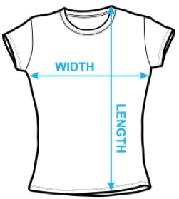
Image Size
Product Details
Our Bella / Canvas t-shirts are made from a 50% cotton / 50% polyester blend and are available in five different sizes. They're stylish, soft, and incredibly comfortable. Machine wash with cold water, and tumble dry on low heat.
Design Details
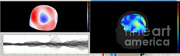
Results of a Magnetoencephalography (MEG) exam carried out on a healthy patient receiving visual stimulation. The curves at bottom left show magnetic... more
Ships Within
1 - 2 business days

Photograph

Canvas Print

Framed Print

Art Print

Poster

Metal Print

Acrylic Print

Wood Print

Greeting Card

iPhone Case

Throw Pillow

Duvet Cover

Shower Curtain

Tote Bag

Round Beach Towel

Zip Pouch

Beach Towel

Weekender Tote Bag

Portable Battery Charger

Bath Towel

Apparel

Coffee Mug

Yoga Mat

Spiral Notebook

Fleece Blanket

Tapestry

Jigsaw Puzzle

Sticker
Results of a Magnetoencephalography (MEG) exam carried out on a healthy patient receiving visual stimulation. The curves at bottom left show magnetic fields both before and during visual stimulation. The right-hand image combines an MRI scan with the MEG. The active areas of the brain during stimulation can be seen (red line). The top left-hand image shows the brain's magnetic flux. MEG detects variations in the brain's magnetic field during various types of cerebral activity. It studies normal and abnormal brain function. Recording the magnetic field produced by neuronal currents requires ultra sensitive sensors called SQUIDs (Superconducting Quantum Interference Device). The 306 sensors spread over 102 areas allow both near and far magnetic fields to be measured, and the brain's deep structures to be 'seen'. Neuroimaging research center, Pitié Salpętričre hospital, Paris, France.
$38.00


There are no comments for Magnetoencephalography #17. Click here to post the first comment.