Use code RJTJFG for 20% off at checkout. Until 5/31/20


by Oliver Meckes EYE OF SCIENCE
$36.00
Color
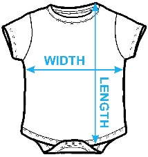
Image Size
Product Details
Our baby onesies are made from 100% pre-shrunk cotton and are available in five different sizes. All baby onesies are machine washable.
Design Details
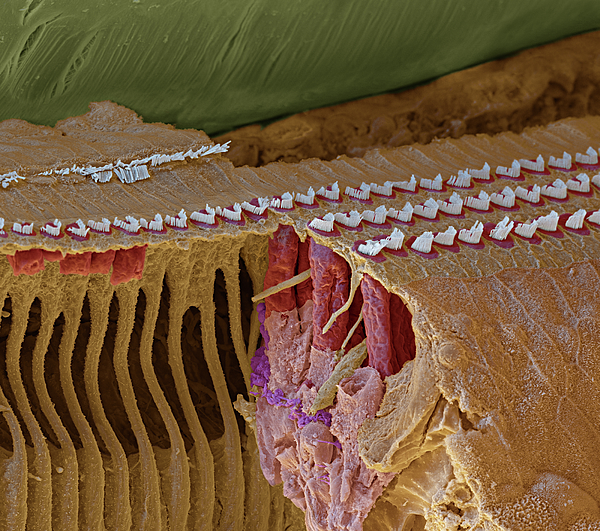
This image shows a part of the cochlea's coil, with the stereovilli of the organ of Corti. It is opened to show the positions of the cells. The three... more
Ships Within
1 - 2 business days

Photograph

Canvas Print

Framed Print

Art Print

Poster

Metal Print

Acrylic Print

Wood Print

Greeting Card

iPhone Case

Throw Pillow

Duvet Cover

Shower Curtain

Tote Bag

Round Beach Towel

Zip Pouch

Beach Towel

Weekender Tote Bag

Portable Battery Charger

Bath Towel

Apparel

Coffee Mug

Yoga Mat

Spiral Notebook

Fleece Blanket

Tapestry

Jigsaw Puzzle

Sticker

Ornament
This image shows a part of the cochlea's coil, with the stereovilli of the organ of Corti. It is opened to show the positions of the cells. The three rows of the outer hair cells (OHC with Stereocilia or Stereovilli, red/white) are clearly visible as the tectorial membrane (greenish), which usually is connected with the tips of the stereocilia has lifted up during preparation. One can see the cylindrical shape of the outer hair cells (red)and the very long extensions of the reinforcing cells (left side). The outermost area right are the "Hensen-cells." Nerve fibers on the base of the outer hair cells are colored violet. The organ of Corti lies in the entire length of the cochlea. Close to the oval window, the hair cells of the organ of Corti are sensitive to high tones, while towards the tip of the cochlea they are sensitive to lower tones. The tectorial membrane is connected to the tips of the hair cells’ stereovilli, which causes these stereovilli to shift and stretch when there is...
$36.00


There are no comments for Cochlea Coil Section, Sem #3. Click here to post the first comment.