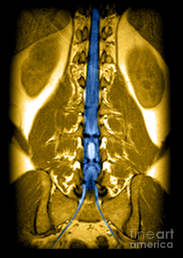
Mri Of Spinal Cord And Nerve Roots #1

by Medical Body Scans
Title
Mri Of Spinal Cord And Nerve Roots #1
Artist
Medical Body Scans
Medium
Photograph - Photograph
Description
This color enhanced coronal (frontal) view of the lower thoracic and lumbar spine beautifully demonstrates the termination of the lower thoracic spinal cord (the conus medullaris) near the thoracolumbar junction. As the spinal cord terminates it tapers to a sharp point which is easily seen in this image.The vertically oriented dark line going through the center of the spinal cord represents the central canal of the spinal cord which normally contains cerebral spinal fluid (CSF). This is normal. In the lower part of the image the S1 nerve roots are beautifully demonstrated as they exit the sacral canal via the sacral neural foramina.
Uploaded
March 7th, 2013
Embed
Share
Comments
There are no comments for Mri Of Spinal Cord And Nerve Roots #1. Click here to post the first comment.


