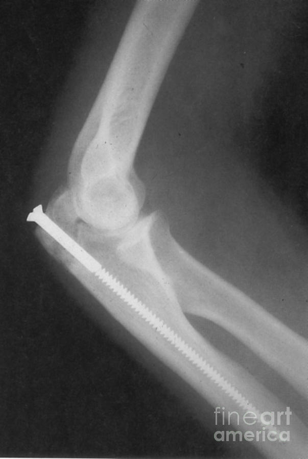
Broken Arm With Metal Pin, X-ray #2

by Science Source
Title
Broken Arm With Metal Pin, X-ray #2
Artist
Science Source
Medium
Photograph - Photograph
Description
X-ray of a broken arm with a metal pin introduced into the marrow cavity in order to strengthen the bone. Wire may also be wound around the bone to assist it to set straight, and metal plates also may assist correct healing in the case of a spiral or comminuted fracture. The surgeon uses anesthesia to relax the muscles and x-ray equipment to help align the bones. A surgeon exposes the fracture to see and manipulate the fragments with special instruments. Then, the bone fragments are securely fixed using some combination of metal wires, pins, screws, rods, and plates. This procedure is called open reduction and internal fixation (ORIF). Metal plates are contoured and fixed to the outside of the bone with screws. Metal rods are inserted from one end of the bone into the marrow cavity. These implants are made of stainless steel, high-strength alloy metal, or titanium. Circa 1980's.
Uploaded
March 14th, 2013
Embed
Share
Comments
There are no comments for Broken Arm With Metal Pin, X-ray #2. Click here to post the first comment.


