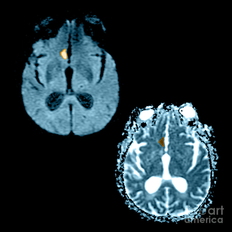
Mri Of Stroke #2

by Medical Body Scans
Title
Mri Of Stroke #2
Artist
Medical Body Scans
Medium
Photograph - Photograph
Description
This composite of 2 axial MRI images reveal the appearance of an acute stroke on diffusion weighted images and the corresponding ADC (apparrent diffusion coefficient) map. The diffusion weighted image in the upper left hand side shows a focal region of increased signal (looks white) in the inferior-medial frontal lobe. The ADC map in the lower right hand side shows that there is restricted (looks brown) diffusion in this region which indicates that the abnormality most likely represents an acute (usually looks like this for 10-14 days) infarct.
Uploaded
March 7th, 2013
Embed
Share
Comments
There are no comments for Mri Of Stroke #2. Click here to post the first comment.


