
The watermark in the lower right corner of the image will not appear on the final print.
Frame
Top Mat

Bottom Mat

Dimensions
Image:
14.00" x 7.00"
Mat Border:
2.00"
Frame Width:
0.88"
Overall:
19.50" x 12.50"
Cancer Cell Death Sequence, Sem #5 Framed Print

by Science Source
Product Details
Cancer Cell Death Sequence, Sem #5 framed print by Science Source. Bring your print to life with hundreds of different frame and mat combinations. Our framed prints are assembled, packaged, and shipped by our expert framing staff and delivered "ready to hang" with pre-attached hanging wire, mounting hooks, and nails.
Design Details
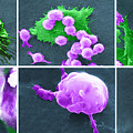
Six-step sequence of the death of a cancer cell. A cancer cell (green) has migrated through the holes of a matrix coated membrane from the top to the... more
Ships Within
3 - 4 business days
Additional Products


Canvas Print

Framed Print

Art Print

Poster

Metal Print

Acrylic Print

Wood Print

Greeting Card

iPhone Case

Throw Pillow

Duvet Cover

Shower Curtain

Tote Bag

Round Beach Towel

Zip Pouch

Beach Towel

Weekender Tote Bag

Portable Battery Charger

Bath Towel

Apparel

Coffee Mug

Yoga Mat

Spiral Notebook

Fleece Blanket

Tapestry

Jigsaw Puzzle

Sticker

Ornament
Framed Print Tags
Comments (0)
Artist's Description
Six-step sequence of the death of a cancer cell. A cancer cell (green) has migrated through the holes of a matrix coated membrane from the top to the bottom, simulating natural migration of an invading cancer cell between, and sometimes through, the vascular endothelium. Notice the spikes or pseudopodia that are characteristic of an invading cancer cell (1). A buffy coat containing macrophages (purple) is added to the bottom of the membrane. A group of macrophages identify the cancer cell as foreign matter and start to stick to the cancer cell, which still has its spikes (2). Macrophages begin to fuse with, and inject its toxins into, the cancer cell. The cell starts rounding up and loses its spikes (3). As the macrophage cell becomes smooth (4). The cancer cell appears lumpy in the last stage before it dies. These lumps are actually the macrophages fused within the cancer cell (5). The cancer cell then loses its morphology, shrinks up and dies (6). Enhancements of BJ4579, BS9471, BS9...
$129.00


There are no comments for Cancer Cell Death Sequence, Sem #5. Click here to post the first comment.